Forensic DNA Laboratory Construction Workflow
Establishing a fully functional forensic DNA laboratory is a complex but structured undertaking. It requires far more than acquiring individual instruments. The core challenge lies in integrating validated workflows, implementing rigorous contamination control, and ensuring long-term operational stability. This guide provides a foundational roadmap for private forensic laboratories, moving from initial concept and spatial design to the seamless integration of a complete DNA analysis workflow. We will cover critical areas including evidence handling, automated DNA extraction using magnetic bead technology, precise PCR amplification, and advanced capillary electrophoresis for genetic analysis. A successful laboratory prioritizes accuracy, efficiency, and adherence to international standards from day one, ensuring that every result, whether for criminal justice or family relationship testing, is legally defensible and scientifically sound. This resource focuses on building a system where each component works in harmony to transform challenging biological evidence into conclusive, high-resolution data.
Initial Laboratory Planning and Infrastructure for Forensic DNA Work
Laboratory Zones & Air Pressure Parameters
| Laboratory Zone | Air Pressure Type | Core Function |
|---|---|---|
| Pre-PCR Area | Positive Pressure | Block external contaminants |
| Amplification Zone | Neutral Pressure | Isolate PCR process |
| Post-PCR Area | Negative Pressure | Contain amplified DNA |
| Evidence Intake | HEPA Filtered | Dry evidence safely |
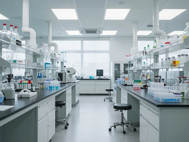
The physical layout and infrastructure of a forensic DNA laboratory are the primary barriers against contamination. A poorly designed space will inevitably lead to sample cross-contamination and false positive results, rendering any expensive equipment useless. The laboratory must follow a unidirectional workflow, meaning evidence moves from the cleanest areas to the dirtiest, with no backtracking. This spatial separation is documented in standards like ISO 18385, which specifically minimizes the risk of human DNA contamination in products used to collect, store, and analyze biological evidence. The facility should be partitioned into dedicated zones for evidence intake, DNA extraction, PCR setup, amplification, and post-amplification analysis such as capillary electrophoresis. Each zone requires independent air handling systems with positive or negative pressure to control the flow of aerosols, a primary vector for DNA contamination in a forensic lab.
Beyond spatial design, the choice of construction materials is critical for decontamination. Surfaces including benchtops, floors, and walls must be non-porous, chemically resistant, and easy to clean. Regular decontamination with specialized DNA remover solutions is essential to degrade any stray genetic material. The laboratory must also be equipped with Class II biological safety cabinets for handling evidence, ensuring both sample protection and operator safety. A dedicated forensic evidence drying cabinet is necessary for processing wet or moist evidence to prevent bacterial and fungal degradation before analysis. Proper storage is equally vital, with upright freezers and refrigerators maintaining the long-term integrity of both evidence and critical reagents. Ignoring these foundational elements is the most common and costly mistake new laboratories make, as retrofitting a space is significantly more difficult than building it correctly from the start.
Unidirectional Workflow and Zonal Separation
Unidirectional workflow is a logistical and procedural concept that physically separates pre- and post-PCR products. The laboratory is divided into three primary zones: a clean or pre-PCR area, an amplification area, and a post-PCR analysis area. Personnel, equipment, and consumables must flow in only one direction. An analyst who enters the post-PCR area cannot return to the pre-PCR area on the same day without a full decontamination procedure. This strict flow prevents amplified DNA products from contaminating evidence samples, a phenomenon that is the leading cause of false positive results in forensic casework. The pre-PCR zone houses evidence examination, DNA extraction, and PCR setup. The amplification zone contains the thermal cyclers, and the post-PCR zone holds the genetic analyzers and other detection instruments.
Each zone requires its own set of dedicated supplies including pipettes, lab coats, gloves, and DNA-free filtered pipette tips. Air pressure differentials are also engineered to support the workflow. The pre-PCR area should maintain positive air pressure to push contaminants out, while the post-PCR area uses negative air pressure to contain any amplified DNA aerosols. Similarly, the evidence drying cabinet should be located in the intake zone and draw air through a HEPA filter to prevent contamination from the general facility. This level of controlled access and environmental separation is not a recommendation but a necessity for any laboratory seeking accreditation from bodies that follow ISO 17025 or similar forensic science standards.
HVAC and Filtration Systems for Contamination Control
Heating, ventilation, and air conditioning systems are not merely for comfort; they are an active defense against molecular contamination. A high-efficiency particulate air filtered system that provides numerous air changes per hour is standard. More importantly, the airflow must be directed from clean zones to dirty zones. For instance, air pressure in the DNA extraction room should be slightly higher than in the hallway, but the hallway's pressure should be higher than the post-PCR room. This creates a cascade that ensures any airborne DNA or aerosols from amplified material cannot drift upstream into the sensitive extraction area. The HVAC system must also maintain stable temperature and humidity levels to ensure the consistent performance of sensitive equipment like capillary electrophoresis genetic analyzers, which use polymers whose viscosity changes with temperature.
Recirculating air within a forensic DNA lab is generally avoided. Most designs use a once-through system where air is exhausted directly to the outside, not back into the main building circulation. The exhaust from the post-PCR area in particular is considered potentially contaminated and must be expelled safely. Regular monitoring of these pressure differentials is part of the laboratory’s quality management system, with alarms installed to alert staff to any failures. This level of environmental control is a significant initial investment, but it directly reduces the rate of inconclusive results and costly re-extractions, providing a return on investment through operational efficiency and judicial reliability.
Essential Support Equipment and Consumables
The daily operation of a forensic DNA lab depends on a variety of smaller support tools and consumables that are easily overlooked during initial planning. Centrifuges of various capacities, including plate centrifuges, palm micro-centrifuges, and multi-rotor centrifuges, are required for sample pelleting and mixing. A digital dry bath incubator is essential for the precise temperature control needed in lysis steps for bone digestion, often running at 56 degrees Celsius overnight with proteinase K. Similarly, vortex mixers and digital orbital shaker mixers are used constantly to ensure homogenous reagent mixtures. A critical but often under-specified piece of equipment is the forensic evidence drying cabinet, which uses unidirectional, HEPA-filtered airflow to dry items like clothing or bedding at room temperature, preventing the degradation of DNA and the growth of mold.
Consumables represent a major recurring cost and source of potential contamination. All plastics including tubes, plates, and tips must be certified DNase, RNase, and human DNA-free. The laboratory should stock various sizes of sterile PCR tubes and plates for different applications. Heat and cold sealing films are used to securely seal plates for PCR, preventing evaporation and cross-well contamination. Disposable shoe covers, powder-free forensic nitrile gloves, and full forensic protective gear including gowns and face masks are worn as the first line of defense against operator-derived DNA. These items are not optional; their consistent use forms the procedural backbone of a contamination-free environment.
The Core Workflow: From Evidence to DNA Extract
The journey from a biological stain to purified DNA is the most critical phase in the forensic process. This workflow involves collecting the sample, isolating cellular material, lysing cells to release DNA, and then purifying that DNA from inhibitors and cellular debris. The primary challenge in forensic science is that evidence samples are often compromised, containing trace amounts of DNA alongside potent inhibitors like heme from blood, humic acid from soil, or indigo dye from denim. An effective workflow must maximize yield while minimizing these co-extracted inhibitors. The choice between manual and automated methods at this stage defines the lab's throughput, consistency, and vulnerability to human error. For a private laboratory, the decision is often driven by case volume and the average difficulty of the samples processed.
Automated systems have become the gold standard for high-throughput and complex forensic casework. These systems are not just about speed; they provide unparalleled consistency and traceability by following the exact same protocol for every sample. An automated 96-channel integrated DNA workstation can process an entire plate of samples, from lysate to purified DNA, in a sealed environment that minimizes hands-on time and contamination risk. For lower volume labs or those processing highly specialized samples like bone, semi-automated or even manual workflows using magnetic or silica bead technology remain viable and cost-effective. The key is matching the level of automation to the specific casework demands, ensuring that the chosen method yields DNA pure enough for the downstream amplification and analysis steps.
Mechanical and Chemical Lysis for Forensic Samples
The first step of DNA extraction is cell lysis, the disruption of cell membranes and nuclear envelopes to release DNA into solution. For soft tissues like blood or buccal swabs, chemical lysis using a detergent-based forensic lysis buffer combined with a proteolytic enzyme is often sufficient. This buffer typically contains sodium dodecyl sulfate to dissolve lipid membranes and a strong chaotropic salt to denature proteins. The enzyme forensic proteinase K is then added to digest these denatured proteins, effectively degrading histones that bind DNA and other cellular structural proteins. This reaction is typically incubated for one to several hours at 56 degrees Celsius with gentle agitation provided by a thermostatic mixing incubator.
Harder samples such as bone, teeth, or even dense fingernails require a more aggressive approach. The physical structure of bone protects DNA within hydroxyapatite crystals. Therefore, mechanical disruption is required before chemical lysis. This is achieved using an automated forensic bone and teeth grinder, which reduces a hard fragment to a fine powder. This powder is then subjected to a prolonged lysis step, often overnight at 56 degrees Celsius, in a buffer containing proteinase K and a reducing agent like dithiothreitol to break disulfide bonds in the tough collagen matrix. The use of a disposable grinding chamber is critical in this step to prevent cross-contamination between bone samples, a notorious challenge in disaster victim identification and missing persons cases. The lysis method must be both efficient to release DNA and gentle enough to prevent shearing it into fragments too small for standard short tandem repeat analysis.
Magnetic Bead Technology vs. Silica Columns
After lysis, the DNA must be separated from the now-chaotic solution containing degraded proteins, lipids, and other cellular components. Two dominant technologies achieve this: silica-based spin columns and magnetic beads. Silica columns rely on the principle that DNA binds to silica in the presence of high concentrations of chaotropic salts. The lysate is passed through a column, the DNA sticks to the silica membrane, and contaminants are washed away. Finally, a low-salt buffer or water is used to elute the purified DNA. This method is simple and effective for clean samples like fresh blood. However, columns can easily clog with particulate matter from degraded samples like soil or bone, leading to low yields. Furthermore, the centrifugation steps required for columns are difficult to automate on a large scale.
Magnetic bead technology uses microscopic particles coated with silica or other ion-exchange surfaces. DNA binds to these beads under specific buffer conditions. A strong magnet is then used to immobilize the beads and their bound DNA against the side of a tube or plate. The liquid containing contaminants is simply aspirated away. The beads are then washed two or three times by resuspending them in wash buffer and reapplying the magnet. Finally, the purified DNA is eluted from the beads. This method is superior for forensic applications because it handles particulate matter well, is easily automated on 96-channel magnetic bead extraction systems, and produces very pure, high-yield DNA. The trade-off is the higher cost of the specialized instruments and the magnetic beads themselves. For a laboratory processing hundreds of trace evidence samples per day, the investment in magnetic bead automation is recouped through reliability and efficiency.
Handling Degraded and Inhibitor-Rich Samples
Forensic DNA extracts are rarely pure. Degraded DNA, fragmented into short pieces, is common in aged or environmentally exposed evidence. Inhibitors that co-extract with DNA can prevent downstream PCR amplification, leading to false-negative results. Common inhibitors include heme compounds from blood, humic and fulvic acids from soil, melanin from hair and skin, and collagen from bone. A robust extraction chemistry must actively remove these substances. Many forensic kits now incorporate specialized wash buffers that are formulated to dissociate these inhibitors from the DNA or the binding matrix. For instance, a high-concentration guanidine hydrochloride wash can remove heme, while a low-concentration alcohol wash can remove polysaccharides.
When working with degraded samples, the choice of binding chemistry becomes crucial. Large DNA fragments bind more readily to silica than small ones. Therefore, protocols designed for degraded DNA often modify the binding conditions, using higher concentrations of polyethylene glycol or other crowding agents to force short fragments onto the beads or column. For extremely challenging samples like formalin-fixed paraffin-embedded tissues or very old skeletal remains, a specialized protocol using a semi-automated large-volume bone DNA kit is required. These kits process a larger starting volume of bone powder lysate to capture a greater absolute amount of DNA, compensating for the high degree of degradation. The final elution volume is also kept small to concentrate the extracted DNA, making it detectable by downstream quantification assays. Success in these cases depends not just on the extraction kit, but on the analyst's understanding of the sample’s taphonomic history and the careful selection of optimized chemistry.
DNA Quantification and Amplification Setup
Optimal DNA Input for PCR Amplification
Knowing the quantity and quality of extracted DNA is mandatory before proceeding to PCR amplification. Forensic DNA laboratories cannot perform blind PCR; an overloaded reaction will produce artifacts like pull-up peaks, while an under-loaded one will result in allele dropout or a partial profile. Quantitative PCR, or real-time PCR, is the standard method because it is highly sensitive and specific for human DNA. Unlike older methods like slot blots, qPCR can discriminate between human and non-human DNA and can even detect the presence of PCR inhibitors in the sample. This is achieved through the use of fluorescent probes that bind specifically to a human target sequence. The instrument measures the cycle at which fluorescence crosses a threshold, which correlates directly to the starting quantity of DNA. Many forensic qPCR kits also include an internal PCR control to flag the presence of inhibitors, providing a critical quality check.
The information from quantification dictates how the PCR amplification step is set up. Forensic labs typically aim to add a specific mass of target DNA, usually between 0.5 and 2.0 nanograms, into the PCR reaction for standard autosomal STR kits. If the extract is pure but low in quantity, the analyst can increase the number of PCR cycles to boost yield. If inhibitors are present, the extract may need to be diluted and re-quantified, as dilution often dilutes the inhibitor enough for PCR to work. This precise setup is non-negotiable for generating clean, interpretable genetic profiles. The entire quantification and setup process must occur in the clean pre-PCR area, using dedicated plate centrifuges to spin down reagents and a biosafety cabinet to maintain a sterile environment. This rigorous upstream process is what enables reliable downstream results from sensitive genetic analyzers.
The Role of Real-Time PCR in Forensics
Real-time PCR technology used in forensic DNA quantification goes beyond simply measuring total human DNA. Advanced multiplex kits can simultaneously quantify both human and male DNA in a single reaction well, a feature essential for sexual assault evidence where a high background of female DNA may obscure a small amount of male DNA. These kits use different fluorescent dyes for the human and Y-chromosome targets. The resulting data allows the analyst to determine if a sample is suitable for Y-STR typing, which targets only the male lineage, or if a differential extraction protocol is required. The human DNA quant PCR kit and Y-chromosome quant PCR kit are examples of targeted assays that provide this specific information.
The sensitivity of modern forensic qPCR systems is remarkable, capable of detecting as little as a few picograms of DNA. However, the quantification system itself must be validated. A standard curve is generated for every run using a DNA standard of known concentration, allowing the instrument to calculate the quantity in the unknown samples. The amplification plots are also analyzed for their shape and slope. A shallow slope can indicate the presence of PCR inhibitors, even if the internal control still amplifies. This sophisticated data provides a risk assessment for each sample, helping the analyst decide whether to proceed with STR amplification or to attempt re-extraction or an alternative method. This quality-driven approach ensures that laboratory resources are not wasted on samples likely to fail, while also preventing the reporting of unreliable results.
Master Mix Assembly and Reaction Setup
Once the DNA is quantified, the PCR setup begins. This step is the most vulnerable to contamination from amplified product or external human DNA. All work is performed in a dedicated PCR setup room equipped with a dead-air box or a dedicated laminar flow hood that is thoroughly decontaminated. The PCR master mix is assembled first. This master mix contains everything needed for the reaction except the template DNA: a thermostable DNA polymerase, deoxynucleotide triphosphates, magnesium chloride, buffer salts, and a set of fluorescently labeled primers targeting specific STR loci. Using a master mix reduces pipetting steps and variability, improving consistency across samples. For forensic work, the DNA polymerase must be highly processive and tolerant of residual inhibitors that survived extraction.
The prepared master mix is then dispensed into the reaction tubes or wells of a 96-well plate, followed by the addition of the purified DNA extract. Each batch of samples must include several controls: a negative control to monitor for contamination, a positive control containing a known DNA standard to ensure the PCR worked, and often an extraction blank that went through the lysis and purification process alongside the evidence. After sealing the plate with a heat-sealing film or a cap strip, it is briefly centrifuged in a plate centrifuge to collect all liquid at the bottom of the wells. The plate is then transferred to the amplification zone. The meticulous nature of this setup directly correlates to the quality of the final genetic profile. Careful pipetting, proper use of sterile consumables, and strict adherence to the lab's anti-contamination protocols are the hallmarks of a professional forensic DNA facility.
Thermal Cycling Parameters for STR Amplification
The PCR amplification of STR markers is performed in a specialized instrument known as a forensic thermal cycler. These instruments are distinct from standard research thermal cyclers in their temperature uniformity and ramp rate specifications. To reliably amplify all loci in a multiplex system, every well in the thermal cycler block must experience the exact same temperature profile. A gradient of even 0.5 degrees Celsius across the block can cause allele dropout or non-specific amplification. High-quality forensic thermal cyclers use Peltier elements and sophisticated algorithms to maintain a uniformity of plus or minus 0.2 degrees Celsius. They also feature heated lids to prevent condensation on the seal, which would change the reaction volume and efficiency.
A typical STR PCR protocol consists of an initial denaturation step at 95 degrees Celsius to separate the DNA double helix, followed by 25 to 30 cycles of denaturation, annealing, and extension. Denaturation at 94-96 degrees Celsius separates the strands. Annealing at 58-62 degrees Celsius allows the primers to bind to their specific target sequences. Extension at 72 degrees Celsius allows the DNA polymerase to synthesize a new strand. The number of cycles is critical; too few cycles produce insufficient product, while too many cycles can lead to the formation of non-specific artifacts. After cycling, a final extension step ensures all strands are fully elongated. The entire process takes approximately two to three hours. The instrument's run log, documenting the thermal profile, is part of the electronic case record. This data is essential for troubleshooting and demonstrates compliance with validated protocols during accreditation inspections.
Capillary Electrophoresis and Genetic Analysis
Capillary Electrophoresis Analysis Steps
The final step in the forensic DNA workflow is separating and detecting the amplified STR fragments. This is accomplished through capillary electrophoresis, a technique that separates DNA fragments by size as they migrate through a thin polymer-filled capillary under an electric field. The genetic analyzer injects the PCR product electrokinetically into the capillary. As fragments of different lengths pass a detector window, a laser excites the fluorescent dyes attached to the primers. The emitted light is captured and converted into an electropherogram, a graph with peaks representing different DNA fragments. The pattern of these peaks at specific loci constitutes the DNA profile, or genetic fingerprint. A capillary electrophoresis genetic analyzer is a highly complex piece of equipment requiring careful maintenance, including the use of running buffer, separation polymers, and periodic replacement of the multichannel capillary array.
This analysis must be performed in the designated post-PCR area, which is isolated from the rest of the laboratory. Before loading onto the genetic analyzer, the PCR product is mixed with formamide and an internal lane standard. The formamide denatures the DNA, ensuring it runs as single strands. The internal lane standard, such as a DNA size standard, is a mix of known DNA fragments labeled with a different color dye than the sample. This standard is run in every capillary alongside the sample, allowing the instrument software to precisely calculate the size of each unknown fragment in base pairs. High-quality reagents like highly deionized formamide and the correct polymer matrix are essential for sharp, well-resolved peaks. Any degradation or contamination in these reagents results in broad peaks or high background noise, rendering the profile unusable.
Fragment Separation and Detection Mechanics
The physics of CE separation is based on the sieving effect of the polymer. The POP-4 or POP-7 polymer forms a network of entangled, long-chain molecules. Under an applied electric field, negatively charged DNA fragments migrate through this network. Smaller fragments weave through the polymer pores more quickly than larger ones. This results in a precise separation based on size, with a resolution of just one base pair possible. The genetic analyzer is typically a multi-capillary instrument, meaning it can run 8, 16, 24, or even 96 samples simultaneously. This parallel processing is what gives forensic laboratories the capacity to handle large casework volumes.
The detection system uses a laser and a charge-coupled device camera. As each labeled fragment passes the detection window, the laser excites the fluorophore, and the camera captures the emission spectrum. Sophisticated software deconvolutes the overlapping spectra from different dyes, assigning each peak a specific color representing a particular dye or STR locus. The data is then analyzed to determine the allele calls. The entire process is controlled by a computer, and the raw data file and analysis parameters are saved permanently. This data trail is part of the electronic laboratory record and is critical for reviewing or re-analyzing results. The precision and automation of modern CE instruments have dramatically reduced the hands-on time for analysis while increasing the resolution and confidence in DNA typing results.
Data Interpretation and Artifact Recognition
Raw data from the genetic analyzer is not a final result. It requires expert human interpretation. The software makes an initial allele call by comparing the size of a sample peak to an allelic ladder, which is a mixture of all the known alleles for a given locus run under the same conditions. However, the analyst must review every peak to confirm it is a true allele and not an artifact. Common artifacts include stutter peaks, which are small peaks one repeat unit smaller than a true allele and caused by the polymerase slipping during PCR. Pull-up peaks occur when an extremely high signal overwhelms the detector, causing the color of that peak to bleed into another dye channel. Dye blobs or spikes are baseline disturbances caused by reagent impurities or electrical fluctuations.
Interpreting low-level DNA samples from trace evidence adds another layer of complexity. In these samples, stochastic effects can cause allele dropout, where one copy of a gene from a heterozygous individual fails to amplify. This can make a mixed DNA sample from two people appear to come from only one person. Laboratories must adhere to strict interpretation guidelines and use statistical frameworks like probabilistic genotyping software to evaluate low-level and mixed DNA profiles. The credibility of the final expert opinion hinges on the laboratory's validated interpretation protocols. The analyst’s job is to apply these rules conservatively, ensuring that no innocent person is falsely implicated by a misinterpreted artifact. This high-stakes analysis is why forensic DNA testing remains a field of highly trained professional scientists, not just automated machinery.
Instrument Maintenance and Performance Verification
A genetic analyzer is a precision instrument that requires a rigorous daily, weekly, and monthly maintenance schedule. Daily maintenance includes cleaning the injection ports and checking the polymer and buffer levels. The capillary regeneration solution is used periodically to recondition the capillary walls, maintaining consistent electrokinetic injection. The conditioning reagent is also run to improve data quality by reducing the buildup of charge on the capillary walls. Before each batch of samples, a spectral calibration is performed to ensure the instrument accurately distinguishes between the different fluorescent dyes. A positive control, typically a known DNA sample with a well-documented profile, is run to verify that the entire system is performing within specification.
Performance is also monitored by examining the resolution, or the ability to separate a 1-base pair difference. This is measured using the peaks from the internal lane standard. The spacing between peaks must meet defined quality thresholds. The signal strength, or RFU, for the control should fall within a validated range. Over time, the multichannel capillary array wears out and must be replaced after a certain number of runs, as the polymer becomes less effective at sieving. Every maintenance action, verification check, and run log is recorded as part of the laboratory's quality system. This meticulous attention to instrument performance is what separates a reliable forensic laboratory from one that produces questionable data. It provides the confidence needed for a court to accept the evidence and for a defendant to trust the result.
Advanced Applications: Degraded DNA and Mixed Samples
Standard forensic workflows are designed for relatively fresh, abundant DNA, but many casework samples are far from ideal. Degraded DNA, broken into short fragments by sunlight, heat, or humidity, is a common challenge. In these cases, standard STR kits that amplify products up to 450 base pairs often fail because the target sequence is simply not intact. To address this, specialized mini-STR kits have been developed. These kits use redesigned primers that bind much closer to the repeat region of the STR locus, generating amplicons that are often less than 250 base pairs. This reduced size dramatically increases the chance of successful amplification from degraded templates. A dedicated degraded DNA analysis solution is often required to systematically address these challenging samples.
Mixed DNA samples, where biological material from two or more individuals is combined, are another major obstacle. A classic example is a sexual assault sample containing a large amount of female epithelial cells and a small amount of male sperm cells. The standard approach is differential extraction, using a two-step lysis process. A gentle lysis buffer breaks open the female epithelial cells, releasing their DNA, while the tough sperm cell walls remain intact. The female DNA is removed, and then a harsher buffer containing a reducing agent like dithiothreitol is used to break open the sperm and release the male DNA. Even with differential extraction, the resulting female DNA background can still be high. In such situations, the laboratory may turn to Y-STR analysis, which targets only the male-specific Y chromosome, completely ignoring the overwhelming female DNA. This capability is a powerful tool for investigating sexual assault cases, especially when the assailant is azoospermic or the sample is degraded. A comprehensive sexual assault forensic evidence workflow integrates both differential extraction and Y-STR testing to maximize information recovery.
Next-Generation Sequencing in Forensic Genetics
While capillary electrophoresis has been the gold standard for decades, next-generation sequencing is emerging as a transformative technology in forensic science. CE measures only the length of an amplified fragment, but NGS can read the actual DNA sequence. This unlocks two major advantages. First, it allows for massively multiplexed assays, where hundreds of genetic markers including STRs, single nucleotide polymorphisms, and insertion-deletions can be analyzed in a single reaction. Second, it provides sequence-level resolution. Two fragments of the same length could have different internal sequences, a phenomenon known as isoalleles. This additional information can help resolve mixed samples and increase the power of discrimination.
A laboratory implementing NGS for casework would require an NGS forensic sequencing system and specialized library preparation kits like a universal forensic panel. The workflow is more complex than CE, involving library preparation, amplification, and bioinformatic analysis. However, the potential benefits for forensic genetics are substantial. For example, the analysis of mitochondrial DNA using NGS is far more comprehensive than traditional Sanger sequencing, which is crucial for analyzing shed hair or very old skeletal remains that lack nuclear DNA. Similarly, forensic genetic genealogy using whole-genome sequencing of SNPs has become a powerful tool for identifying unknown suspects through familial DNA database searching, though it raises significant privacy and ethical considerations. The integration of NGS into a forensic lab is a major strategic decision that redefines the scope of what is testable, pushing the boundaries of evidence analysis far beyond traditional STR typing.
Investigative Genetic Genealogy and DNA Databases
Beyond individual casework, forensic DNA laboratories support large-scale identification projects and national security initiatives. The maintenance of a DNA database, such as a national criminal offender index, requires high-throughput processing. Laboratories dedicated to this function rely on fully automated 96-channel integrated workstations that can extract, amplify, and analyze thousands of samples per week with minimal human intervention. The consistency and reliability of automated systems are paramount for populating these databases, where errors can have cascading consequences. Similarly, disaster victim identification following a mass fatality event demands a coordinated response. The laboratory must process hundreds of reference samples from family members and compare them to post-mortem samples from the deceased. A structured disaster victim identification protocol is essential for managing the logistics and maintaining accuracy under time pressure.
In these high-volume scenarios, the choice of consumables directly impacts efficiency. The use of buccal collection cards or FTA blood collection cards simplifies sample storage and transport, as the chemical matrix on the card preserves the DNA and inactivates pathogens. The DNA can often be used directly in a PCR reaction after a simple punch and wash, reducing extraction time. The laboratory's information management system becomes as critical as its wet-lab equipment. A Laboratory Information Management System tracks every sample from intake through analysis, managing the workflow and ensuring chain of custody. For missing persons and unidentified remains cases, a dedicated missing persons DNA identification solution is required, which often combines autosomal STRs, Y-STRs, and mitochondrial DNA analysis to increase the chances of a match across distant relatives. The role of the forensic lab in this context extends from pure science to humanitarian action, providing answers to families and closure to communities.
Quality Assurance, Accreditation, and Legal Compliance
The ultimate currency of a forensic DNA laboratory is not its equipment or throughput, but the trustworthiness of its results. Every procedure, from evidence receipt to data analysis, must be documented, validated, and auditable. This is the domain of Quality Assurance. The laboratory must operate under a quality management system that conforms to international standards, the most important being ISO 17025, which specifies the general requirements for the competence of testing and calibration laboratories. For forensic DNA, an additional document, ISO 18385, provides specific requirements for minimizing the risk of human DNA contamination in the products used to collect and analyze biological evidence. Achieving accreditation against these standards is a rigorous, expensive, but non-negotiable process for any laboratory that wishes its results to be admissible in a court of law.
Compliance is not a one-time event but a continuous process of monitoring, validation, and corrective action. All equipment, including thermal cyclers, genetic analyzers, and centrifuges, must undergo regular calibration and performance verification. Every new batch of reagents, from extraction kits to PCR master mixes, must be tested and qualified before use in casework. Key personnel must participate in proficiency testing, where they analyze unknown samples and submit their results for external review. The culture of the lab must be one of transparency and continuous improvement. An accredited lab maintains a comprehensive document control system, with standard operating procedures for every task. All deviations from these procedures are documented as non-conformances and investigated. This rigorous infrastructure is what defends the laboratory's work during a Daubert or Frye hearing, ensuring the judge and jury can rely on the scientific evidence presented.
Validation of Forensic DNA Workflows
Before a new method or instrument can be used on actual evidence, it must undergo a formal validation. This is a scientifically rigorous process that defines the performance characteristics and limitations of the system. For a DNA extraction kit, this involves testing it on a wide range of forensic samples including blood, saliva, semen, and touch DNA, both fresh and degraded. The studies measure the yield and purity of the DNA and test the extract for inhibitors. For a genetic analyzer, validation includes assessing its precision, accuracy, and sensitivity. The laboratory must determine the minimum amount of DNA that can be reliably detected and typed, known as the stochastic threshold. All of this data is compiled into a validation report that becomes a permanent part of the laboratory's quality records.
There are two types of validation: developmental and internal. Developmental validation is performed by the manufacturer to demonstrate that a new kit or instrument works as intended. Internal validation is performed by the end-user laboratory to demonstrate that the method works in their specific environment, with their personnel, and on their caseload of samples. For example, a laboratory adopting a new autosomal STR casework kit would run hundreds of known samples, mixtures, and degraded samples to establish their own interpretation guidelines. They would also carry out contamination studies to ensure their physical layout and procedures are adequate. This local validation is a critical link between a manufacturer's claim and real-world forensic performance. A court will often ask whether the specific technique used was properly validated by the laboratory, making the validation report a key piece of evidence in itself. A complete forensic DNA workflow solution includes pre-validated methods that simplify the internal validation burden while maintaining rigorous scientific standards.
Chain of Custody and Evidence Integrity
The scientific validity of a DNA result is meaningless if the evidence's integrity in the legal chain cannot be proven. The chain of custody is a chronological documentation that records the seizure, custody, control, transfer, analysis, and disposition of physical or electronic evidence. Every person who handles the evidence, from the crime scene investigator to the forensic analyst to the clerk who stores it, must document the transfer in a log. This log must be complete and without gaps. Any break in the chain can be used by the defense to argue that the evidence was tampered with or contaminated, potentially leading to its exclusion from trial.
The laboratory’s role in the chain is to create and maintain meticulous records. Upon evidence intake, the lab assigns a unique laboratory number. Every sample tube, extract, and PCR plate is labeled with this number and a unique identifier. Electronic records track which analyst accessed which sample, for how long, and what they did. Secure refrigerators and freezers with restricted access and continuous temperature monitoring store the biological evidence. The turnkey forensic DNA lab design includes a secure evidence storage area. The analyst’s notes, instrument data files, and the final report are all part of the case file. This comprehensive paper and electronic trail is the backbone of the legal admissibility of DNA evidence. Without it, the most sophisticated scientific analysis is legally useless.
Developing a Laboratory Quality Manual
The quality manual is the foundational document for a forensic DNA laboratory. It describes the laboratory's quality system, including its organizational structure, policies, and procedures. The manual defines the scope of activities, documents the management's commitment to quality, and outlines the responsibilities of each role from the laboratory director to the technical staff. For a new lab, developing this manual in consultation with an accreditation body is the first major milestone. The manual is a living document, updated as procedures change or new equipment is added. It serves as the primary training resource for new employees and the main reference document for auditors.
A well-constructed quality manual also details the laboratory's procedures for handling non-conformances, customer complaints, and corrective actions. It specifies the document control process, ensuring that only the most current version of any SOP is in use. It also describes the internal audit schedule, where laboratory staff review their own compliance with the quality system. For a private forensic lab, the quality manual is not merely an internal tool; it is a public-facing credential that demonstrates competence and reliability to clients including law enforcement agencies, private attorneys, and courts. Investing the time and expertise to build a comprehensive quality manual is as important as purchasing any piece of analytical equipment.
Conclusion: Building a Sustainable Forensic DNA Laboratory
Key Equipment Maintenance Schedule
| Equipment | Maintenance Frequency |
|---|---|
| Genetic Analyzer | Daily Inspection + Monthly Calibration |
| Thermal Cycler | Weekly Performance Check |
| HVAC System | 24h Pressure Monitoring |
| Safety Cabinets | Monthly Certification |
Constructing a forensic DNA laboratory is a multi-faceted project that demands equal attention to physical infrastructure, analytical equipment, and quality systems. The process begins with a unidirectional laboratory design incorporating positive and negative pressure zones to control contamination. It proceeds through the careful selection of extraction, quantification, amplification, and analysis platforms, with a consistent emphasis on validated performance with forensic samples. The operational heart of the lab lies in its standard operating procedures, its trained personnel, and its commitment to continuous quality improvement through internal and external audits.
Sustainability requires a long-term view of operating costs including consumables, equipment service contracts, and ongoing training. The decision between manual, semi-automated, and fully automated workflows should balance initial capital expenditure against long-term labor costs and throughput requirements. A laboratory that successfully integrates physical separation, validated analytical processes, and a rigorous quality management system gains the trust of the judicial system and the community it serves. The ultimate measure of success is not the sophistication of the instruments, but the reliability of the results and the confidence they inspire in the pursuit of truth and justice. For those ready to undertake this challenge, the path is well-defined, the standards are clear, and the tools are available.