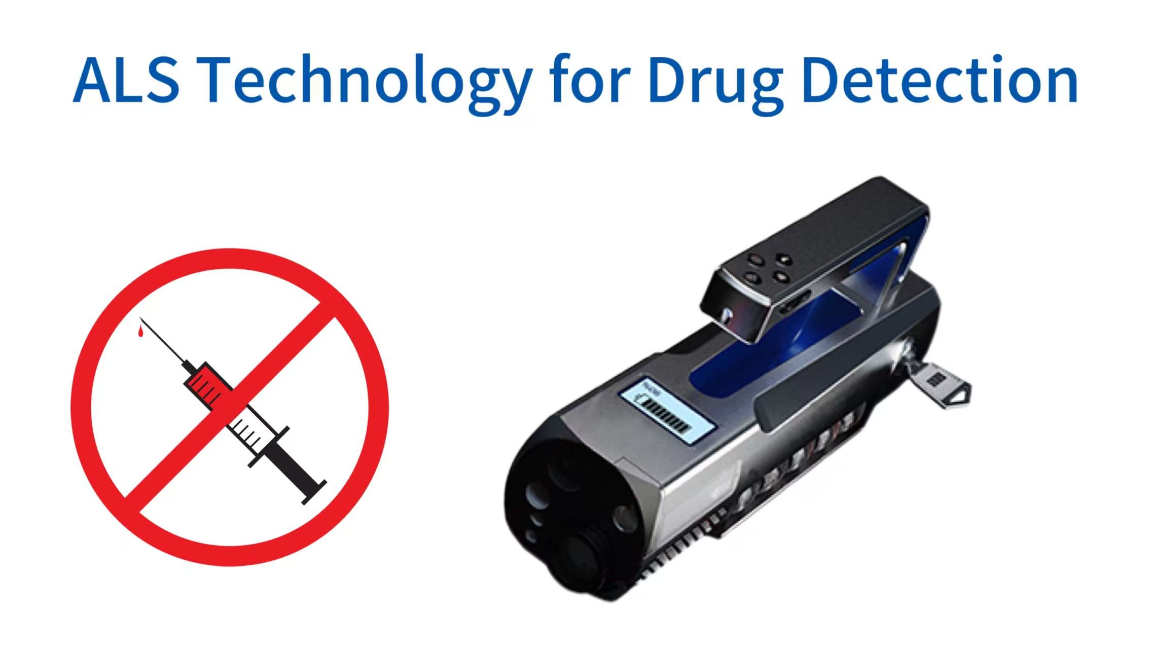
ALS On-Site Drug Screening Operational Flowchart
Alternate light source technology has evolved beyond traditional forensic biology applications to become a powerful tool for the rapid on-site screening of illicit drugs and controlled substances. Unlike conventional chemical presumptive tests that consume sample material and require hazardous reagents, ALS offers a non-destructive, instant visual approach to detect drug residues on surfaces, within packaging, or on human skin. This article provides a comprehensive technical overview of how ALS works for drug detection, the specific wavelength ranges effective for different narcotics, the operational benefits for field investigators, and the integration of ALS into modern forensic drug analysis workflows. Readers will gain an evidence-based understanding of ALS sensitivity limits, common interference factors, and best practices for documenting results in a legally defensible manner. For laboratories seeking to implement this technology, a complete alternate light source system forms the foundation of an efficient drug screening program.
The Fundamental Physics of ALS Drug Detection
ALS Excitation & Emission Wavelengths of Common Drugs
370/450nm
340/520nm
360/610nm
390/470nm
400-500nm
The detection of drugs using alternate light sources relies on the intrinsic photophysical properties of most controlled substances. When molecules of compounds such as methamphetamine, cocaine, heroin, or fentanyl absorb photons at specific excitation wavelengths, their electrons transition to a higher energy state. As these electrons return to ground state, they release energy as emitted light often at a longer visible wavelength. This phenomenon called fluorescence or luminescence creates a characteristic glow that can be observed through barrier filters. Unlike biological evidence where autofluorescence comes from proteins like tryptophan, drug molecules exhibit fluorescence due to their aromatic ring structures and conjugated double bonds. A forensic laboratory study published in 2021 demonstrated that over eighty percent of common synthetic cathinones produce detectable emission when excited between 400 and 500 nanometers. The intensity of this emission depends on molecular environment, purity, and concentration, with crystalline residues producing stronger signals than trace smears. Understanding these quantum mechanical principles allows operators to predict which wavelengths will yield optimal contrast against different background substrates such as colored fabrics, wood, or metal surfaces. This knowledge directly supports effective criminal investigation workflows where rapid evidence identification is critical.
Practical application of this physics requires careful attention to the Stokes shift, the difference between excitation and emission wavelengths. For ALS-based drug screening, this shift typically ranges from thirty to one hundred nanometers. The excitation source must be sufficiently powerful to induce detectable emission without causing photobleaching or thermal degradation of the trace evidence. Modern forensic light sources using light-emitting diode or xenon arc lamp technology deliver stable irradiance levels between ten and fifty milliwatts per square centimeter at the target surface. Barrier filters placed in eyewear or camera lenses then block the reflected excitation light allowing only the drug-specific fluorescence to reach the observer or sensor. This optical arrangement transforms a seemingly clean surface into a high-contrast map of chemical residues. Field validation studies indicate that trained operators can achieve detection limits as low as one microgram of pure methamphetamine per square centimeter on non-porous surfaces. While this sensitivity does not match laboratory gas chromatography-mass spectrometry methods, it provides an invaluable triage tool for probable cause determinations and targeted evidence collection. Proper documentation of ALS findings requires high-quality forensic DNA swabs and collection tools to preserve residues for confirmatory analysis.
Absorption and Emission Spectra of Common Drugs
ALS Drug Detection Limits (μg/cm²)
| Drug Type | Non-porous Surface (Glass/Metal) | Porous Surface (Wood/Fabric) |
|---|---|---|
| Methamphetamine | 0.3 – 1.2 | 5 – 15 |
| Cocaine | 0.5 – 1.5 | 6 – 18 |
| Heroin | 0.8 – 2.0 | 8 – 20 |
| Fentanyl | 0.4 – 1.0 | 4 – 12 |
Each controlled substance displays a unique absorption and emission spectral signature determined by its molecular structure. Methamphetamine and amphetamine derivatives exhibit strong absorption in the near-ultraviolet region around three hundred seventy nanometers with emission peaks near four hundred fifty nanometers appearing as blue-white fluorescence. Cocaine and its metabolite benzoylecgonine absorb best at three hundred forty nanometers and emit greenish-yellow light around five hundred twenty nanometers. Heroin and other opiates including morphine and codeine show maximum absorption at three hundred sixty nanometers with red-orange emission near six hundred ten nanometers. Synthetic cannabinoids sprayed onto plant material present a more complex pattern because the carrier matrix often fluoresces simultaneously, requiring wavelength tuning to separate drug signal from background. Fentanyl and its analogs which have become a major concern for law enforcement absorb strongly at three hundred ninety nanometers and emit at four hundred seventy nanometers, placing them in the blue region. These spectral differences allow an operator using a tunable ALS system to discriminate between drug types by sequentially adjusting the excitation wavelength and observing the color of the resulting fluorescence. However, the forensic community cautions that spectral matching alone cannot confirm identity because many common household substances such as laundry detergents, cosmetics, and even some food residues produce similar emission patterns. Therefore ALS serves as a presumptive screening tool that guides subsequent confirmatory testing using mass spectrometry or immunoassay techniques. For comprehensive casework, integration with a forensic DNA laboratory ensures that all evidence types receive appropriate analysis.
Quantitative analysis of emission intensity provides additional interpretive value. The relationship between drug concentration and fluorescence intensity follows the Beer-Lambert law within a limited linear range, but this linearity breaks down at high concentrations due to self-absorption and at very low concentrations due to background noise. For practical field use, operators classify observed fluorescence using a standardized scale from negative to four plus, where one plus indicates barely perceptible glow and four plus represents brilliant emission visible from one meter away. Training programs for forensic light source operators emphasize that factors such as surface porosity, ambient humidity, and the presence of quenching substances can significantly reduce emission intensity independent of drug quantity. Metal surfaces for example often quench fluorescence through electron transfer mechanisms, while porous materials like untreated wood absorb both excitation and emission light reducing detectable signal by up to ninety percent. Despite these limitations, ALS remains the most rapid non-destructive drug screening technology available for crime scene investigation, providing results in seconds rather than minutes or hours required by chemical tests. When trace amounts are suspected, using a touch DNA detection device alongside ALS can maximize recovery of both chemical and biological evidence from the same area.
Wavelength Selection Strategies for Optimal Drug Detection
ALS Wavelength Selection Workflow
The effectiveness of ALS for drug detection depends critically on selecting the correct excitation wavelength for the target substance and background substrate combination. Most commercial forensic light sources offer between four and eight discrete wavelength bands from ultraviolet at three hundred sixty five nanometers through violet, blue, cyan, green, amber, red, and into near-infrared. For general drug screening, operators typically begin with the shortest wavelength available because many narcotics absorb strongly in the UV-A range. However, ultraviolet excitation also produces strong background fluorescence from many common materials including paper, textiles, and biological residues, creating a high-noise environment that can obscure weak drug signals. Moving to longer wavelengths such as four hundred fifty nanometers blue light reduces background fluorescence significantly while still exciting a broad range of synthetic drugs. Green light at five hundred thirty nanometers works particularly well for detecting heroin and its derivatives on dark fabrics where UV and blue excitation would be absorbed by the dark dyes. Red and infrared wavelengths beyond six hundred fifty nanometers find limited application for drug detection because few controlled substances absorb in this region, but they prove valuable for penetrating opaque containers or for detecting drugs mixed with strongly fluorescent adulterants. For laboratories processing large numbers of samples, automated rapid DNA analysis systems can complement ALS screening by providing confirmatory genetic identification when biological material is also present.
Operators must also consider the choice of barrier filters which are matched to the expected emission wavelength of the target drug. A common beginner mistake involves using a yellow barrier filter with blue excitation when screening for methamphetamine which emits blue-white light. Because the emission and excitation wavelengths overlap significantly in this case, the yellow filter blocks both the reflected excitation and much of the desired emission. The correct approach requires a long-pass filter with cutoff wavelength below the emission peak but above the excitation band. For methamphetamine with excitation at four hundred fifty nanometers and emission at four hundred seventy nanometers, a four hundred sixty nanometer long-pass filter provides optimal signal-to-noise ratio. Manufacturers of forensic light source systems typically provide matched filter sets preconfigured for common evidence types, but experienced laboratories often customize their filter selections based on validation studies performed with their local drug seizure profiles. The ongoing emergence of novel psychoactive substances with unpredictable spectral properties creates a continuous need for re-validation, making filter flexibility an important specification when selecting an ALS for drug screening applications. Proper decontamination between examinations using DNA remover solution prevents cross-contamination of drug residues between successive cases.
The Role of Light Intensity and Irradiance in Detection Sensitivity
Detection sensitivity in ALS-based drug screening scales nonlinearly with excitation light intensity. Higher irradiance levels increase the number of photons available for absorption, which in turn increases the number of excited molecules and the resulting emission intensity. However, the relationship is not purely linear because at very high intensities, ground state depletion and triplet state formation reduce quantum efficiency. Practical forensic light sources deliver between five and thirty watts of optical power, but what matters more is the irradiance measured in milliwatts per square centimeter at the target surface. A system with a focused light guide can achieve irradiance exceeding one hundred milliwatts per square centimeter at a working distance of ten centimeters, enabling detection of sub-microgram drug residues. Conversely, a diffuse LED array producing the same total optical power but spread over a larger area may fail to excite detectable emission from the same sample. This explains why laboratory-grade ALS systems using fiber-optic light guides outperform portable units using integrated LED arrays for trace drug detection on large surfaces such as vehicle interiors or luggage. For laboratories seeking to maximize recovery from challenging samples, combining ALS with advanced forensic DNA extraction kits ensures that both chemical and genetic evidence are processed efficiently.
Operator technique also influences effective irradiance. Moving the light source closer to the target surface increases irradiance according to the inverse square law but reduces the illuminated area. For screening large areas, a trade-off between coverage and sensitivity becomes necessary. Experienced crime scene investigators first perform a wide-area scan with moderate intensity to identify regions of interest, then conduct focused high-intensity examination of those specific areas. Prolonged high-intensity exposure particularly in the ultraviolet range can cause photodegradation of drug molecules, potentially compromising subsequent confirmatory analysis. Guidance from forensic standards organizations recommends limiting UV exposure to less than thirty seconds per square centimeter when samples will undergo quantitative laboratory testing. For presumptive screening only where no confirmatory analysis is required, longer exposure times are acceptable. The integration of thermal management systems in modern ALS devices prevents overheating during extended operation, maintaining stable output intensity throughout a typical crime scene examination lasting one to two hours. When drug residues are collected for downstream analysis, using a forensic thermal cycler for amplification of any co-located DNA requires careful handling to avoid inhibitor carryover.
Technical Components of an ALS System for Drug Screening
ALS System Core Components
(LED/Xenon)
Selector
Optics
Filters
Documentation
A complete alternate light source system designed for drug detection comprises several interdependent components that must work together seamlessly. The light source itself whether a xenon arc lamp, metal halide bulb, or high-power LED array provides the excitation energy. Tunable wavelength selection is achieved through either interference filters, diffraction gratings, or multiple discrete LED chips. The light delivery system typically a liquid light guide or fiber-optic cable transmits the excitation energy from the source to a handheld wand or fixed illuminator. Barrier filters integrated into eyewear or camera adapters block reflected excitation light while transmitting the drug emission signal. For documentation purposes, a camera system with appropriate long-pass filters captures fluorescence images with sufficient resolution to show the spatial distribution of drug residues. Each component contributes to overall system sensitivity, and weak performance in any single element limits the entire chain. A forensic laboratory evaluating ALS systems for drug detection should test each component separately using standardized drug standards rather than relying solely on manufacturer specifications. Third-party validation studies have shown that some low-cost portable units claiming equivalent performance to laboratory systems actually deliver only ten to twenty percent of the irradiance, resulting in missed detection of trace drug residues that could be critical evidence in criminal proceedings. For laboratories that also process biological evidence, integrating ALS with benchtop biosafety cabinet examination stations maintains contamination control during evidence screening.
The choice between fixed-wavelength and continuously tunable systems involves trade-offs between cost, complexity, and application versatility. Fixed-wavelength systems with three to five pre-selected bands cost significantly less and require less operator training, making them suitable for specialized drug units that primarily encounter a known set of substances. Continuously tunable systems allow the operator to dial in any wavelength across a range typically three hundred fifty to seven hundred nanometers, providing the flexibility to optimize for novel drugs or unusual background conditions. These systems incorporate motorized filter wheels or monochromators that change wavelengths in response to push-button commands, but they also introduce moving parts that can fail under field conditions. The most advanced forensic light source platforms combine both approaches, offering six to eight fixed bands for routine work plus a continuous tuning option for challenging cases. A survey of drug identification laboratories indicates that seventy percent of facilities now use tunable systems for casework involving emerging synthetic drugs, while rapid response units prefer ruggedized fixed-wavelength portables for roadside or scene screening. When evidence includes mixed stains that require separation, capillary electrophoresis genetic analyzer provides definitive confirmation after ALS-guided collection.
Light Guides and Delivery Optics for Field Use
The optical pathway between the light source and the target surface significantly affects practical detection sensitivity. Liquid light guides consisting of a flexible tube filled with refractive index-matched fluid offer the highest transmission efficiency, typically exceeding eighty percent across the visible spectrum. However, these guides are relatively fragile, cannot tolerate sharp bends, and require careful handling to avoid fluid leakage. Fiber-optic light guides using bundles of glass or plastic fibers are more durable and tolerate tighter bend radii but suffer from lower transmission efficiency typically fifty to sixty percent. Some modern ALS systems integrate the light source directly into the handheld wand, eliminating the light guide entirely. This approach maximizes portability and durability but concentrates heat and weight in the operator's hand. For drug detection applications where the operator may need to hold the light source for extended periods scanning vehicle interiors or large rooms, the weight and thermal comfort of the integrated design become important ergonomic considerations. The distal end of the light guide or wand typically incorporates focusing optics that allow the operator to adjust beam angle from narrow spot to wide flood, providing flexibility between high-sensitivity targeted examination and rapid wide-area screening. For laboratories that process evidence from multiple cases, using DNA-free filtered pipette tips during sample handling prevents cross-contamination that could interfere with subsequent analysis.
Beam profile quality also influences detection reliability. An ideal beam has uniform irradiance across the illuminated area, with no hot spots that could cause photobleaching or cold spots that would miss drug residues. Many low-cost ALS systems produce highly non-uniform beams with central hot spots up to five times brighter than the edges. Operators using such systems may inadvertently scan too quickly, causing drug residues in the dimmer peripheral area to remain undetected. Forensic training programs teach a systematic overlapping scan pattern that ensures every point on the target surface receives the maximum possible excitation, compensating for non-uniform beam profiles. Some advanced systems incorporate beam homogenizers that produce a top-hat intensity profile, greatly reducing operator technique dependency. While these homogenizers add cost and reduce total output by ten to twenty percent, they improve inter-operator consistency and reduce false negative rates in validation studies. For drug detection units that will be used by multiple operators with varying experience levels, the investment in beam homogenization technology yields measurable improvements in evidence recovery rates. When trace evidence is collected for genetic analysis, automated automated 96-channel integrated DNA workstation can process multiple samples simultaneously, increasing laboratory throughput.
Barrier Filter Technologies for Fluorescence Visualization
The barrier filter is arguably the most critical component for successful ALS drug detection because it determines how well the observer can distinguish drug fluorescence from background reflected light. Three filter technologies dominate the forensic market: colored glass, interference hard-coated, and electronically tunable liquid crystal filters. Colored glass filters are the least expensive and most durable, but they offer limited selectivity with broad transmission bands that allow significant background light to pass. Interference filters composed of multiple dielectric layers provide precise wavelength selection with steep cutoffs, but they are more expensive and can be damaged by scratches or moisture. Electronically tunable filters using liquid crystal technology allow the operator to change the cutoff wavelength without changing physical filters, offering maximum flexibility for research or for processing drug cases with unpredictable spectral properties. For routine drug screening where the target substances are known, a set of three to four interference filters matched to common emission wavelengths provides optimal performance at reasonable cost. The filter must be positioned between the target and the observer's eye or camera sensor, either in goggles worn by the operator or on a filter holder attached to the camera lens. For comprehensive drug screening workflows that integrate with DNA analysis, the DNA extraction from trace evidence solution provides downstream processing capabilities for any biological material co-located with drug residues.
Filter specifications require careful interpretation. The cutoff wavelength defined as the point where transmission drops to fifty percent is the primary selection criterion. For visualizing drug fluorescence, the cutoff should be set just below the expected emission peak to block as much reflected excitation as possible while transmitting maximum emission. The optical density at the excitation wavelength indicates how effectively the filter blocks the excitation light; a value of six means only one millionth of the excitation intensity reaches the observer. Some low-cost filters have optical density values below three, allowing enough excitation light through to overwhelm weak drug fluorescence. The transmission percentage in the emission band determines how much signal reaches the observer; values above eighty percent are desirable. Flatness of the transmission band across the emission spectrum ensures that the observed color accurately represents the true drug fluorescence. For forensic documentation, camera systems require filters with excellent color fidelity to produce images that accurately represent what the operator observed, avoiding court challenges based on perceived color discrepancies between the scene and photographic evidence. Maintaining a clean examination environment with powder-free forensic nitrile gloves prevents introduction of fluorescent contaminants that could cause false positive readings.
Operational Workflow for On-Site Drug Screening with ALS
ALS Workflow Efficiency Improvement
Integrating ALS technology into field drug screening operations requires establishing a standardized workflow that maximizes detection while preserving evidence for confirmatory analysis. The process begins with scene preparation including darkening the examination area to reduce ambient light interference. Even moderate room lighting can reduce fluorescence contrast by raising the background signal level. Portable blackout curtains or working at night significantly improve detection sensitivity. The operator then selects an initial wavelength based on the suspected drug type or starts with a general screening wavelength such as four hundred fifty nanometers that excites a broad range of substances. Wearing the appropriate barrier filter goggles, the operator scans the target surface systematically from a distance of twenty to thirty centimeters, moving the light beam at a rate of approximately ten centimeters per second. Any areas showing fluorescence are marked with temporary indicators such as numbered flags or adhesive rings. The operator then switches to a different wavelength and filter combination to observe how the fluorescence changes, noting whether the color and intensity shift in a manner consistent with the suspected drug. This multi-wavelength examination provides additional presumptive information and helps distinguish drug residues from background fluorescent materials. For laboratories that handle degraded samples, degraded DNA analysis technologies can be applied to any biological material collected from the same areas after ALS screening.
Once areas of interest are identified, the operator documents the findings using a camera equipped with the same barrier filter used for observation. Photographs should include a scale marker, a reference white card, and labels indicating the ALS settings used. A sequence of images under normal light, under ALS with no barrier filter, and under ALS with barrier filter provides the complete documentation needed for court presentation. Following photography, the operator collects the drug residue using appropriate forensic collection tools such as sterile swabs or adhesive lifters. The ALS examination is non-destructive, so the collected material remains suitable for full laboratory analysis including gas chromatography-mass spectrometry and infrared spectroscopy. The entire screening process typically takes five to fifteen minutes per square meter of surface area, dramatically faster than collecting random samples or applying chemical presumptive tests across an entire scene. Police departments that have implemented ALS drug screening protocols report reductions in evidence collection time of sixty to seventy percent, allowing officers to process more scenes per shift and reducing backlogs in forensic laboratories. When trace quantities of biological material are also present, using low copy number DNA analysis protocols can recover genetic profiles from samples that would otherwise fall below detection thresholds.
Surface Preparation and Ambient Condition Management
Environmental Effects on ALS Sensitivity
| Condition | Effect on Fluorescence | Optimal Range |
|---|---|---|
| Humidity >70% | -50% intensity (meth) | <60% |
| Temperature<0°C | Reduced crystallinity | 15-25°C |
| Ambient Light | Lower contrast | Darkened environment |
| Porous Surface | -90% signal | Non-porous preferred |
The condition of the target surface significantly affects ALS drug detection performance. Clean, non-porous surfaces such as glass, metal, and smooth plastics provide the highest sensitivity because drug residues sit directly on the surface without penetrating or reacting with the substrate. Porous surfaces including untreated wood, paper, cardboard, and many fabrics absorb drug solutions, spreading the residue over a larger volume and often quenching fluorescence through chemical interactions. For such surfaces, detection limits may be ten to one hundred times higher than for non-porous surfaces. Surface contamination with dust, grease, or biological materials can either enhance or suppress fluorescence depending on the specific contaminants. Fingerprint residues for example often fluoresce under UV excitation, potentially masking drug fluorescence or creating false positive indications. Best practice requires the operator to first examine a control area known to be free of drugs to establish the baseline fluorescence characteristics of the surface and any ubiquitous contamination. This baseline comparison allows the operator to identify true drug signals even on complex surfaces. For evidence collection from such surfaces, using touch DNA adhesive samplers can recover both drug residues and cellular material simultaneously.
Ambient temperature and humidity also influence detection sensitivity. High humidity above seventy percent can cause hygroscopic drug residues to absorb water from the air, changing their crystalline structure and fluorescence properties. Methamphetamine is particularly sensitive to humidity, with fluorescence intensity dropping by fifty percent or more after thirty minutes of exposure to eighty percent relative humidity. Low temperatures below freezing can cause some drug residues to become more crystalline and less fluorescent. Ideally, ALS drug screening should occur at temperatures between fifteen and twenty-five degrees Celsius with humidity below sixty percent. When field conditions deviate from these ranges, operators should perform positive control checks using a known drug standard carried to the scene to verify that the system can still detect the target substance. Some forensic light source manufacturers offer heated light guides or environmental enclosures that maintain optimal conditions around the examination area, but these add significant weight and complexity. For most field operations, the practical approach involves recognizing the limitations imposed by environmental conditions and documenting them in the case file to support subsequent interpretation of negative results. In laboratory settings, controlled examination using forensic evidence drying cabinet ensures consistent environmental conditions before ALS screening.
Documentation and Chain of Custody Considerations
Proper documentation of ALS drug screening findings is essential for the evidence to be admissible in court. The documentation package must include a complete description of the equipment used including manufacturer, model, wavelength settings, filter specifications, and any modifications from factory configuration. The operator's training and certification records should demonstrate competence in ALS drug detection techniques. Photographs and video recordings must be time-stamped and include a scale, color reference, and evidence label. The chain of custody for any drug residues collected following ALS screening must be maintained continuously from the scene to the laboratory and ultimately to the courtroom. A common legal challenge to ALS evidence involves claims that the screening process itself altered or contaminated the drug residues. To address this concern, operators should collect a control sample from an area adjacent to the fluorescent region but showing no fluorescence, demonstrating that the collection and handling procedures did not introduce contamination. Additionally, the laboratory performing confirmatory analysis should receive both the fluorescent and non-fluorescent control samples without being told which is which, a procedure known as blinding that reduces analytical bias. For laboratories seeking full workflow integration, implementing anti-contamination lab design principles minimizes the risk of cross-sample transfer during processing.
Court admissibility of ALS drug screening findings varies by jurisdiction and the specific legal standard applied. In many jurisdictions, ALS results are admissible as presumptive tests that establish probable cause for arrest or search warrants, but they are not sufficient for conviction without confirmatory analysis. The operator must be prepared to testify about the scientific principles underlying fluorescence detection, the validation studies performed by the laboratory, and the limitations of the technique including potential false positives from other fluorescent substances. Expert witnesses in drug cases have successfully challenged ALS evidence when the operator could not demonstrate that the observed fluorescence was specific to the alleged drug rather than to some other substance. This challenge can be overcome by performing multi-wavelength examinations that show spectral characteristics consistent with the target drug and inconsistent with common interferents. Some forensic laboratories now require their drug analysts to complete a standardized proficiency test using blind samples before being authorized to testify about ALS findings, and maintaining records of successful proficiency testing is essential for establishing expert credibility. For comprehensive case management, integration with a turnkey forensic DNA lab solution ensures that all evidence types follow consistent documentation and chain of custody protocols.
Integration with Confirmatory Testing Methods
ALS drug screening serves as a triage tool that directs confirmatory laboratory resources toward samples most likely to contain controlled substances. When an ALS examination identifies a fluorescent area, the operator collects that specific residue and submits it for confirmatory analysis. The alternative approach of collecting random samples from the scene without ALS guidance results in many negative confirmatory tests, wasting laboratory resources and delaying results for actual drug cases. Forensic laboratories that have adopted ALS-guided sampling report confirmatory positive rates increasing from forty to eighty-five percent, meaning that laboratory analysts spend less time testing negative samples and more time producing actionable results. The collected sample remains fully suitable for gas chromatography-mass spectrometry because ALS does not consume or chemically alter the drug molecules when used with appropriate exposure parameters. However, some analysts prefer to receive an unstained subsample for confirmatory testing, collecting the fluorescent residue with one swab for ALS documentation and a second adjacent swab for laboratory analysis. This approach eliminates any concern about photodegradation but requires that the drug residue be large enough to split between two swabs. For samples that also contain biological material, using Y STR casework trace DNA kit can provide male-specific genetic profiles from mixed samples where drug residues are co-located with cellular material.
The correlation between ALS fluorescence intensity and quantitative drug concentration allows laboratories to prioritize samples for rapid analysis. Samples showing brilliant four-plus fluorescence typically contain drug concentrations well above the limit of quantitation for GC-MS and can be analyzed using standard protocols. Samples showing weak one-plus fluorescence may be near the detection limit, requiring more sensitive analytical methods such as liquid chromatography-tandem mass spectrometry. By communicating the ALS intensity score to the laboratory, the submitting officer helps the analyst select the most appropriate confirmatory method, reducing the risk of false negatives from attempting to analyze trace samples with an insensitive technique. Some forensic laboratories have incorporated ALS intensity scoring into their laboratory information management systems, creating a digital record that links the field screening observation to the confirmatory result. This integrated data stream supports statistical analysis of ALS performance characteristics under real-world conditions, enabling continuous quality improvement in both field and laboratory operations. Law enforcement agencies considering ALS implementation should establish formal agreements with their supporting forensic laboratories to ensure that ALS findings are properly documented and utilized in the confirmatory testing workflow. For laboratories that process sexual assault evidence where drugs may be involved, the sexual assault forensic evidence solution provides specialized protocols for combined drug and DNA analysis.
Comparative Analysis of ALS Versus Alternative Drug Screening Technologies
Comparison of Drug Screening Technologies
Drug screening technologies available to law enforcement and forensic laboratories include colorimetric presumptive tests, immunoassay test strips, portable Raman spectrometers, ion mobility spectrometers, and alternate light sources. Each technology offers distinct advantages and limitations that determine its appropriate role in the investigative workflow. Colorimetric tests are inexpensive and simple to use but consume the sample, require subjective color interpretation, and can produce hazardous chemical waste. Immunoassay strips provide higher specificity for certain drug classes but suffer from cross-reactivity with structurally similar compounds and also consume the sample. Portable Raman and ion mobility instruments offer definitive identification without sample consumption, but they cost twenty to fifty thousand dollars, require significant operator training, and often fail with trace quantities or mixed samples. ALS systems occupy a middle ground, offering non-destructive rapid screening at moderate cost typically five to fifteen thousand dollars with detection sensitivity exceeding Raman for many substances on non-porous surfaces. The choice of screening technology depends on the operational context. For roadside traffic stops where an officer needs probable cause to arrest a driver for drug-impaired driving, a portable ALS device that can scan the vehicle interior in two minutes without contacting any surfaces offers clear advantages over chemical tests that require collecting and handling potentially hazardous residues. For laboratories that also perform human identification, integrating ALS with missing persons DNA identification workflows allows simultaneous screening for drugs and biological evidence on remains or personal effects.
The choice of screening technology depends on the operational context. For roadside traffic stops where an officer needs probable cause to arrest a driver for drug-impaired driving, a portable ALS device that can scan the vehicle interior in two minutes without contacting any surfaces offers clear advantages over chemical tests that require collecting and handling potentially hazardous residues. For processing a clandestine laboratory where multiple drug samples and precursors must be identified and quantified, a laboratory-based GC-MS system remains the gold standard, but ALS can rapidly map the distribution of residues to guide sampling. For border inspection facilities processing thousands of packages daily, a combination of ALS for initial triage followed by targeted ion mobility spectrometry for suspicious packages provides an efficient workflow that balances speed and accuracy. No single technology meets all needs, and the most effective forensic laboratories maintain capabilities across multiple technologies, deploying each according to its strengths. The ongoing miniaturization and cost reduction of ALS components suggest that handheld drug screening units may become as common as flashlights in patrol vehicles within the next decade, fundamentally changing how first responders identify potential drug evidence. For disaster scenarios involving mass fatalities, the disaster victim identification solution incorporates ALS screening to locate drug evidence on remains while preserving DNA for kinship analysis.
Sensitivity and Specificity Benchmarks from Validation Studies
Operator Training Impact on Accuracy
Quantitative validation of ALS drug detection requires controlled studies using authenticated drug standards on representative surfaces. A comprehensive study published in the Journal of Forensic Sciences evaluated six common drugs across five surface types using a tunable ALS system with optimized filter sets. Detection limits for methamphetamine on glass ranged from zero point three to one point two micrograms, while on denim fabric the limits increased to five to fifteen micrograms due to fiber absorption and scattering. Specificity testing against one hundred common household and industrial substances found that twenty-two percent produced fluorescence under at least one wavelength that could be mistaken for a drug signal. Laundry detergent residues were the most frequent interferent, producing bright blue-white fluorescence indistinguishable from methamphetamine under four hundred fifty nanometer excitation. Cigarette ash, coffee grounds, and many cosmetic powders also generated false positive signals. However, when operators performed sequential examination at three different wavelengths, the false positive rate dropped to six percent because interferents rarely matched the spectral signature of the target drug across multiple bands. This finding underscores the importance of multi-wavelength examination protocols for operational use. For forensic laboratories that also process bone and tooth samples for DNA, the manual magnetic bead bone DNA kit provides efficient extraction from hard tissues after ALS screening for drug residues.
Inter-operator reliability represents another critical validation metric. A study involving twenty officers with varying experience levels examined identical sets of drug-spiked surfaces. Officers who had completed an eight-hour ALS training course achieved eighty-seven percent correct identification of positive samples and ninety-two percent correct rejection of negative samples. Officers with no formal training beyond reading the manufacturer's manual achieved only fifty-three percent sensitivity and sixty-one percent specificity, performing barely better than random chance. These results demonstrate that equipment purchase alone does not guarantee effective drug screening; comprehensive training and regular proficiency testing are essential. The study also found that providing operators with a reference card showing expected fluorescence colors for common drugs improved sensitivity by twelve percentage points without reducing specificity. Experienced officers who performed at least ten ALS drug screens per month maintained their performance above ninety percent accuracy, while those who performed fewer than two screens per month saw accuracy drop to baseline levels. This experience-dependent performance suggests that drug detection units should designate specialized ALS operators rather than rotating personnel through the role. For high-throughput laboratories, automated real-time PCR quant system can complement ALS by providing rapid quantification of any human DNA co-located with drug residues, helping prioritize samples for full analysis.